
|
- 品牌:OminimAbs
- 产地:上海
- 型号:毫升
- 货号:电询
- 发布日期: 2024-08-26
- 更新日期: 2024-10-09
| 产地 | 上海 |
| 品牌 | OminimAbs |
| 保存条件 | |
| 货号 | 电询 |
| 用途 | 实验研究 |
| 应用范围 | 科研实验 |
| 抗原来源 | 电询 |
| 保质期 | 2年 |
| 抗体名 | 电询 |
| 是否单克隆 | |
| 克隆性 | 否 |
| 靶点 | 电询 |
| 适应物种 | 电询 |
| 形态 | 电询 |
| 宿主 | 电询 |
| 标记物 | 电询 |
| 包装规格 | 毫升 |
| 亚型 | 电询 |
| 标识物 | 电询 |
| 浓度 | 电询% |
| 免疫原 | 电询 |
| 是否进口 | 否 |
Product Profile
Product Name
Anti-CD142 antibody
Antibody Type
Primary Antibodies
Immunogen
Polypeptide
Key Feature
Clonality
Polyclonal
Isotype
IgG
Host Species
Rabbit
Tested
Application
ELISAICC/IFWB
WB:1:200-1:2000
ICC/IF:1:100-1:500
Species
Reactivity
HumanMouseRat
Concentration
1 mg/ml
Purification
Protein A
Target Information
Gene Symbol
F3
Gene Synonyms
TF
TFA
CD142
F3
tissue factor
Hide
Gene Full Name
coagulation factor III, tissue factor
Gene Summary
This gene encodes coagulation factor III
which is a cell surface glycoprotein. This factor enables cells to initiate
the blood coagulation cascades, and it functions as the high-affinity
receptor for the coagulation factor VII. The resulting complex provides a
catalytic event that is responsible for initiation of the coagulation
protease cascades by specific limited proteolysis. Unlike the other cofactors
of these protease cascades, which circulate as nonfunctional precursors, this
factor is a potent initiator that is fully functional when expressed on cell
surfaces, for example, on monocytes. There are 3 distinct domains of this
factor: extracellular, transmembrane, and cytoplasmic. Platelets and
monocytes have been shown to express this coagulation factor under
procoagulatory and proinflammatory stimuli, and a major role in
HIV-associated coagulopathy has been described. Platelet-dependent monocyte
expression of coagulation factor III has been described to be associated with
Coronavirus Disease 2019 (COVID-19) severity and mortality. This protein is
the only one in the coagulation pathway for which a congenital deficiency has
not been described. Alternate splicing results in multiple transcript
variants.[provided by RefSeq, Aug 2020]
Hide
Molecular
Weight(MW)
33 kDa(Observed:50
kDa)
Source
Rabbit
Cellular
Localizatio
Plasma Membrane,Extracellular region or
secreted
Application
·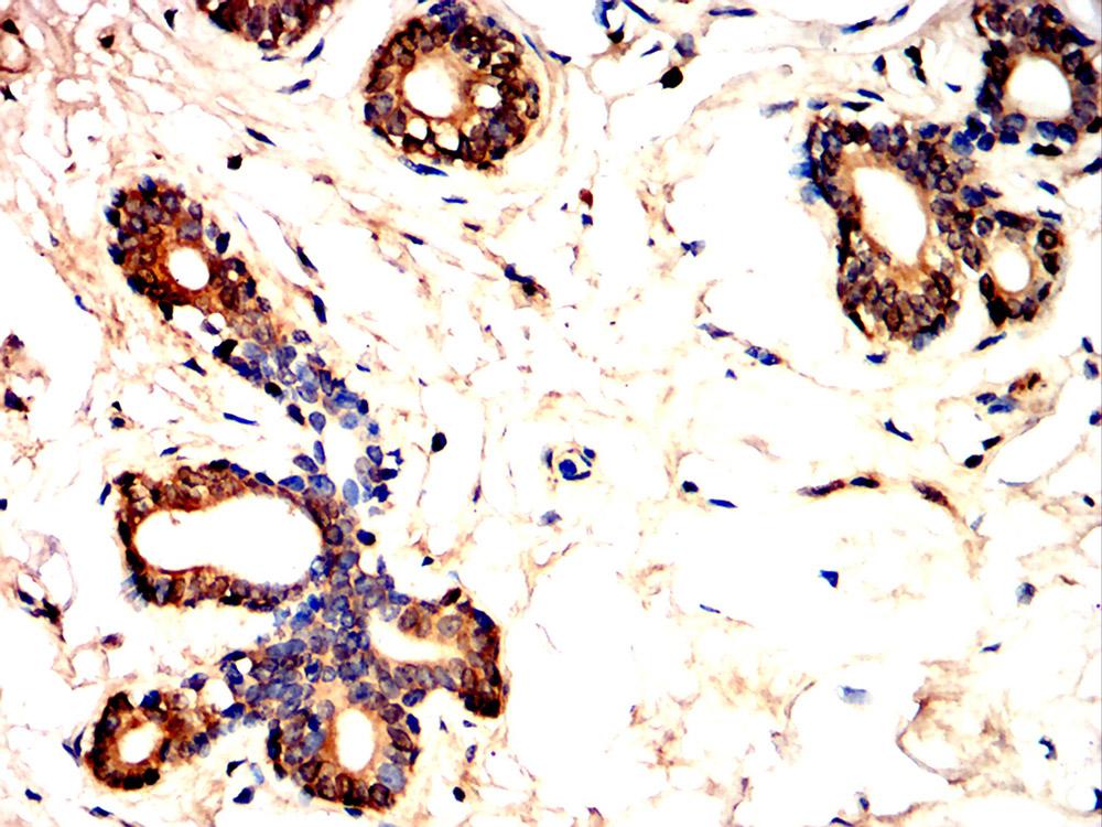
WB
Western blot
analysis using CD142 antibody against Raji (1), HepG2 (2), Hela (3), HUVEC (4)
cell lysate.
·
ICC/IF
Immunofluorescence
analysis of Hela cells using CD142 antibody (green). Blue: DAPI fluorescent DNA
dye. Red: Actin filaments have been labeled with Alexa Fluor- 555 phalloidin.
Application
Notes
WB:1:200-1:2000
ICC/IF:1:100-1:500
Additional Information
Form
Liquid
Storage
Instruction
Shipped at 4°C. Store at +4°C short term
(1-2 weeks). Store at -20°C long term. Avoid freeze / thaw cycle.
Storage Buffer
Purified antibody in PBS with 0.05%
sodium azide.
Product Profile
Product Name
Anti-CD142 antibody
Antibody Type
Primary Antibodies
Immunogen
Polypeptide
Key Feature
Clonality
Polyclonal
Isotype
IgG
Host Species
Rabbit
Tested
Application
ELISAICC/IFWB
WB:1:200-1:2000
Species
Reactivity
HumanMouseRat
Concentration
1 mg/ml
Purification
Protein A
Target Information
Gene Symbol
F3
Gene Synonyms
TF
Hide
Gene Full Name
coagulation factor III, tissue factor
Gene Summary
This gene encodes coagulation factor III
which is a cell surface glycoprotein. This factor enables cells to initiate
the blood coagulation cascades, and it functions as the high-affinity
receptor for the coagulation factor VII. The resulting complex provides a
catalytic event that is responsible for initiation of the coagulation
protease cascades by specific limited proteolysis. Unlike the other cofactors
of these protease cascades, which circulate as nonfunctional precursors, this
factor is a potent initiator that is fully functional when expressed on cell
surfaces, for example, on monocytes. There are 3 distinct domains of this
factor: extracellular, transmembrane, and cytoplasmic. Platelets and
monocytes have been shown to express this coagulation factor under
procoagulatory and proinflammatory stimuli, and a major role in
HIV-associated coagulopathy has been described. Platelet-dependent monocyte
expression of coagulation factor III has been described to be associated with
Coronavirus Disease 2019 (COVID-19) severity and mortality. This protein is
the only one in the coagulation pathway for which a congenital deficiency has
not been described. Alternate splicing results in multiple transcript
variants.[provided by RefSeq, Aug 2020]
Hide
Molecular
Weight(MW)
33 kDa(Observed:50
kDa)
Source
Rabbit
Cellular
Localizatio
Plasma Membrane,Extracellular region or
secreted
Application
ICC/IF:1:100-1:500
TFA
CD142
F3
tissue factor
·
WB
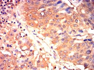
Western blot analysis using CD142 antibody against Raji (1), HepG2 (2), Hela (3), HUVEC (4) cell lysate.
·
ICC/IF
Immunofluorescence analysis of Hela cells using CD142 antibody (green). Blue: DAPI fluorescent DNA dye. Red: Actin filaments have been labeled with Alexa Fluor- 555 phalloidin.
|
Application Notes |
WB:1:200-1:2000 |
Additional Information
|
Form |
Liquid |
|
Storage Instruction |
Shipped at 4°C. Store at +4°C short term (1-2 weeks). Store at -20°C long term. Avoid freeze / thaw cycle. |
|
Storage Buffer |
Purified antibody in PBS with 0.05% sodium azide. |

-
中秋迎好礼 中秋迎好礼 随着国产3A大作《黑神话:悟空》的面世,现我司推出活动。 从即日起至2024年9月18日,购买ELISA试剂盒满五盒,即可申领《黑神话:悟空》数字标准版兑换码。 ... VIEW DETAILS
-
化学实验室守则小分享 一、实验前 在进行化学实验前,学生必须完成预习工作,了解实验的目的、步骤和注意事项。进入实验室前,应穿着适当的防护装备,如实验服、护目镜、实验手套等。此外,学生应提... VIEW DETAILS
-
犬肾素(REN)ELISA试剂盒在宠物健康监测中的重要作用 随着宠物健康管理的日益受重视,犬肾素(REN)ELISA试剂盒在宠物健康监测中发挥着不可或缺的作用。 犬肾素作为一种关键的激素,其水平的变化能够反映犬只肾脏的功能状态。而犬肾素(REN)ELISA试... VIEW DETAILS
-
上海笃玛生物科技有限公司2024年春节放假通知 尊敬的新老客户: 您好! 2024年春节即将到来,上海笃玛生物科技有限公司全体员工提前恭祝广大新老客户,新春快乐、生意兴隆、万事如意!非常感谢您长期以来对我们工作的理解... VIEW DETAILS
-
科研基础小知识分享,细胞传代 一、什么是细胞传代? 细胞传代是细胞培养过程中的一项基本技术,是指将培养的细胞在一定的条件下进行分离、再培养的过程。传代过程中,细胞会逐渐生长,并填满整个培养容器,因此需要进... VIEW DETAILS
-
ELISA样品制备&收集指南 一、引言 酶联免疫吸附试验(ELISA)是一种广泛应用于生物学和医学研究的免疫分析方法。该方法通过将抗原或抗体固相化在载体表面,实现对目标分子的捕获和检测。在进行ELISA实验之前,... VIEW DETAILS

- 手机:15214367449
- Q Q: