
|
- 品牌:OminimAbs
- 产地:上海
- 型号:毫升
- 货号:OM642008
- 发布日期: 2024-08-23
- 更新日期: 2024-10-09
| 产地 | 上海 |
| 品牌 | OminimAbs |
| 保存条件 | |
| 货号 | OM642008 |
| 用途 | 实验研究 |
| 应用范围 | 科研实验 |
| 抗原来源 | 电询 |
| 保质期 | 2年 |
| 抗体名 | 电询 |
| 是否单克隆 | 否 |
| 克隆性 | 否 |
| 靶点 | 电询 |
| 适应物种 | 电询 |
| 形态 | 电询 |
| 宿主 | 电询 |
| 标记物 | 电询 |
| 包装规格 | 毫升 |
| 亚型 | 电询 |
| 标识物 | 电询 |
| 浓度 | 电询% |
| 免疫原 | 电询 |
| 是否进口 | 否 |
Product Profile
Product Name
Anti-TLR7 antibody
Antibody Type
Primary Antibodies
Immunogen
Polypeptide
Key Feature
Clonality
Polyclonal
Isotype
IgG
Host Species
Rabbit
Tested
Application
ELISAICC/IFIHCWB
WB:1:200-1:2000
IHC:1:200-1:1000
ICC/IF:1:100-1:500
Species
Reactivity
HumanMouseRat
Concentration
1 mg/ml
Purification
Protein A
Target Information
Gene Symbol
TLR7
Gene Synonyms
IMD74
TLR7-like
Gene Full Name
toll like receptor 7
Gene Summary
The protein encoded by this gene is a
member of the Toll-like receptor (TLR) family which plays a fundamental role
in pathogen recognition and activation of innate immunity. TLRs are highly
conserved from Drosophila to humans and share structural and functional
similarities. The human TLR family comprises 11 members. They recognize
pathogen-associated molecular patterns (PAMPs) that are expressed on
infectious agents, and mediate the production of cytokines necessary for the
development of effective immunity. For the recognition of structural
components in foreign microorganisms, the various TLRs exhibit different
patterns of expression as well; in this way for example, TLR-3, -7, and -8
are essential in the recognition of single-stranded RNA viruses. TLR7 senses
single-stranded RNA oligonucleotides containing guanosine- and uridine-rich
sequences from RNA viruses, a recognition occuring in the endosomes of
plasmacytoid dendritic cells and B cells. This gene is predominantly
expressed in lung, placenta, and spleen, and is phylogenetically related and
lies in close proximity to another family member, TLR8, on chromosome X.
[provided by RefSeq, Aug 2020]
Hide
Molecular
Weight(MW)
121 KDa
Source
Rabbit
Cellular
Localizatio
Endoplasmic reticulum membrane. Endosome.
Lysosome. Cytoplasmic vesicle > phagosome
Application
· 
WB
Western blot
analysis using TLR7 antibody against PANC-1 (1) cell lysate.
· 
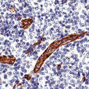
IHC
Immunohistochemical
analysis of paraffin-embedded thyroid carcinoma tissues using TLR7 antibody
with DAB staining.
· 
ICC/IF
Immunofluorescence
analysis of A549 cells using TLR7 antibody (green). Blue: DAPI fluorescent DNA
dye. Red: Actin filaments have been labeled with Alexa Fluor- 555 phalloidin.
Application
Notes
WB:1:200-1:2000
IHC:1:200-1:1000
ICC/IF:1:100-1:500
Additional Information
Form
Liquid
Storage
Instruction
Shipped at 4°C. Store at +4°C short term
(1-2 weeks). Store at -20°C long term. Avoid freeze / thaw cycle.
Storage Buffer
Purified antibody in PBS with 0.05%
sodium azide.
Product Profile
Product Name
Anti-TLR7 antibody
Antibody Type
Primary Antibodies
Immunogen
Polypeptide
Key Feature
Clonality
Polyclonal
Isotype
IgG
Host Species
Rabbit
Tested
Application
ELISAICC/IFIHCWB
WB:1:200-1:2000
IHC:1:200-1:1000
ICC/IF:1:100-1:500
Species
Reactivity
HumanMouseRat
Concentration
1 mg/ml
Purification
Protein A
Target Information
Gene Symbol
TLR7
Gene Synonyms
IMD74
TLR7-like
Gene Full Name
toll like receptor 7
Gene Summary
The protein encoded by this gene is a
member of the Toll-like receptor (TLR) family which plays a fundamental role
in pathogen recognition and activation of innate immunity. TLRs are highly
conserved from Drosophila to humans and share structural and functional
similarities. The human TLR family comprises 11 members. They recognize
pathogen-associated molecular patterns (PAMPs) that are expressed on
infectious agents, and mediate the production of cytokines necessary for the
development of effective immunity. For the recognition of structural
components in foreign microorganisms, the various TLRs exhibit different
patterns of expression as well; in this way for example, TLR-3, -7, and -8
are essential in the recognition of single-stranded RNA viruses. TLR7 senses
single-stranded RNA oligonucleotides containing guanosine- and uridine-rich
sequences from RNA viruses, a recognition occuring in the endosomes of
plasmacytoid dendritic cells and B cells. This gene is predominantly
expressed in lung, placenta, and spleen, and is phylogenetically related and
lies in close proximity to another family member, TLR8, on chromosome X.
[provided by RefSeq, Aug 2020]
Hide
Molecular
Weight(MW)
121 KDa
Source
Rabbit
Cellular
Localizatio
Endoplasmic reticulum membrane. Endosome.
Lysosome. Cytoplasmic vesicle > phagosome
Application
·
WB
Western blot
analysis using TLR7 antibody against PANC-1 (1) cell lysate.
·
IHC
Immunohistochemical
analysis of paraffin-embedded thyroid carcinoma tissues using TLR7 antibody
with DAB staining.
·
ICC/IF
Immunofluorescence
analysis of A549 cells using TLR7 antibody (green). Blue: DAPI fluorescent DNA
dye. Red: Actin filaments have been labeled with Alexa Fluor- 555 phalloidin.
Application
Notes
WB:1:200-1:2000
IHC:1:200-1:1000
ICC/IF:1:100-1:500
Additional Information
Form
Liquid
Storage
Instruction
Shipped at 4°C. Store at +4°C short term
(1-2 weeks). Store at -20°C long term. Avoid freeze / thaw cycle.
Storage Buffer
Purified antibody in PBS with 0.05%
sodium azide.
Product Profile
Product Name
Anti-TLR7 antibody
Antibody Type
Primary Antibodies
Immunogen
Polypeptide
Key Feature
Clonality
Polyclonal
Isotype
IgG
Host Species
Rabbit
Tested
Application
ELISAICC/IFIHCWB
WB:1:200-1:2000
Species
Reactivity
HumanMouseRat
Concentration
1 mg/ml
Purification
Protein A
Target Information
Gene Symbol
TLR7
Gene Synonyms
IMD74
Gene Full Name
toll like receptor 7
Gene Summary
The protein encoded by this gene is a
member of the Toll-like receptor (TLR) family which plays a fundamental role
in pathogen recognition and activation of innate immunity. TLRs are highly
conserved from Drosophila to humans and share structural and functional
similarities. The human TLR family comprises 11 members. They recognize
pathogen-associated molecular patterns (PAMPs) that are expressed on
infectious agents, and mediate the production of cytokines necessary for the
development of effective immunity. For the recognition of structural
components in foreign microorganisms, the various TLRs exhibit different
patterns of expression as well; in this way for example, TLR-3, -7, and -8
are essential in the recognition of single-stranded RNA viruses. TLR7 senses
single-stranded RNA oligonucleotides containing guanosine- and uridine-rich
sequences from RNA viruses, a recognition occuring in the endosomes of
plasmacytoid dendritic cells and B cells. This gene is predominantly
expressed in lung, placenta, and spleen, and is phylogenetically related and
lies in close proximity to another family member, TLR8, on chromosome X.
[provided by RefSeq, Aug 2020]
Hide
Molecular
Weight(MW)
121 KDa
Source
Rabbit
Cellular
Localizatio
Endoplasmic reticulum membrane. Endosome.
Lysosome. Cytoplasmic vesicle > phagosome
Application
IHC:1:200-1:1000
ICC/IF:1:100-1:500
TLR7-like
·
WB
Western blot analysis using TLR7 antibody against PANC-1 (1) cell lysate.
·
IHC
Immunohistochemical analysis of paraffin-embedded thyroid carcinoma tissues using TLR7 antibody with DAB staining.
·
ICC/IF
Immunofluorescence analysis of A549 cells using TLR7 antibody (green). Blue: DAPI fluorescent DNA dye. Red: Actin filaments have been labeled with Alexa Fluor- 555 phalloidin.
|
Application Notes |
WB:1:200-1:2000 |
Additional Information
|
Form |
Liquid |
|
Storage Instruction |
Shipped at 4°C. Store at +4°C short term (1-2 weeks). Store at -20°C long term. Avoid freeze / thaw cycle. |
|
Storage Buffer |
Purified antibody in PBS with 0.05% sodium azide. |
-
中秋迎好礼 中秋迎好礼 随着国产3A大作《黑神话:悟空》的面世,现我司推出活动。 从即日起至2024年9月18日,购买ELISA试剂盒满五盒,即可申领《黑神话:悟空》数字标准版兑换码。 ... VIEW DETAILS
-
化学实验室守则小分享 一、实验前 在进行化学实验前,学生必须完成预习工作,了解实验的目的、步骤和注意事项。进入实验室前,应穿着适当的防护装备,如实验服、护目镜、实验手套等。此外,学生应提... VIEW DETAILS
-
犬肾素(REN)ELISA试剂盒在宠物健康监测中的重要作用 随着宠物健康管理的日益受重视,犬肾素(REN)ELISA试剂盒在宠物健康监测中发挥着不可或缺的作用。 犬肾素作为一种关键的激素,其水平的变化能够反映犬只肾脏的功能状态。而犬肾素(REN)ELISA试... VIEW DETAILS
-
上海笃玛生物科技有限公司2024年春节放假通知 尊敬的新老客户: 您好! 2024年春节即将到来,上海笃玛生物科技有限公司全体员工提前恭祝广大新老客户,新春快乐、生意兴隆、万事如意!非常感谢您长期以来对我们工作的理解... VIEW DETAILS
-
科研基础小知识分享,细胞传代 一、什么是细胞传代? 细胞传代是细胞培养过程中的一项基本技术,是指将培养的细胞在一定的条件下进行分离、再培养的过程。传代过程中,细胞会逐渐生长,并填满整个培养容器,因此需要进... VIEW DETAILS
-
ELISA样品制备&收集指南 一、引言 酶联免疫吸附试验(ELISA)是一种广泛应用于生物学和医学研究的免疫分析方法。该方法通过将抗原或抗体固相化在载体表面,实现对目标分子的捕获和检测。在进行ELISA实验之前,... VIEW DETAILS

- 手机:15214367449
- Q Q: